What is microscopic brain tumour surgery?
A brain tumour is an abnormal growth of cells inside the head. Some tumours are benign (not cancer) and grow slowly. Others are malignant (cancer) and can grow faster. Both types can cause problems by pressing on the brain.
Read moreShow less
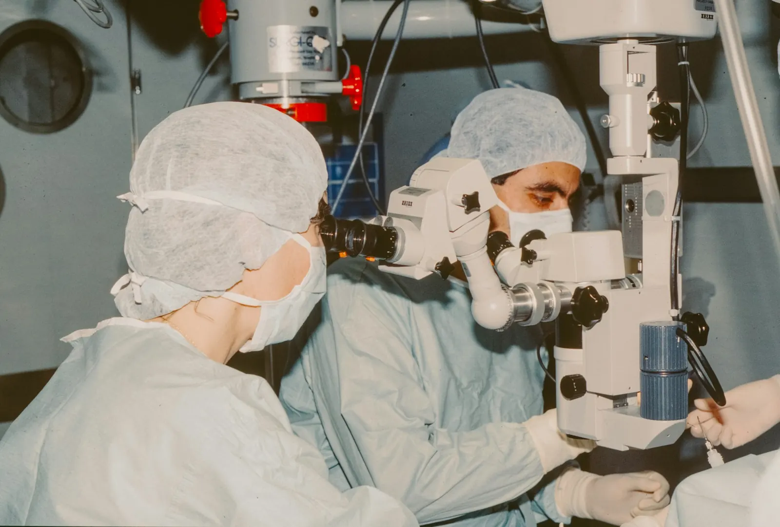
In most brain tumour operations, the surgeon opens a small window in the skull. This is called a craniotomy. An operating microscope gives a bright, magnified view so the tumour can be separated from normal brain tissue. Tools such as neuronavigation (a computer guidance system, like GPS for the brain, that uses your scans) may be used to plan the safest path.
Sometimes only a small piece of the tumour is taken out for testing. This is called a biopsy. It is often done when the tumour is deep, in a sensitive area, or likely to be treated mainly with radiotherapy or chemotherapy. The tissue report helps decide the next steps.
Who may need it
Surgery is one option, not the only one. It is decided after an examination and a careful look at your scans, and non-surgical treatment is discussed first where it is suitable.
How it is done
- 1
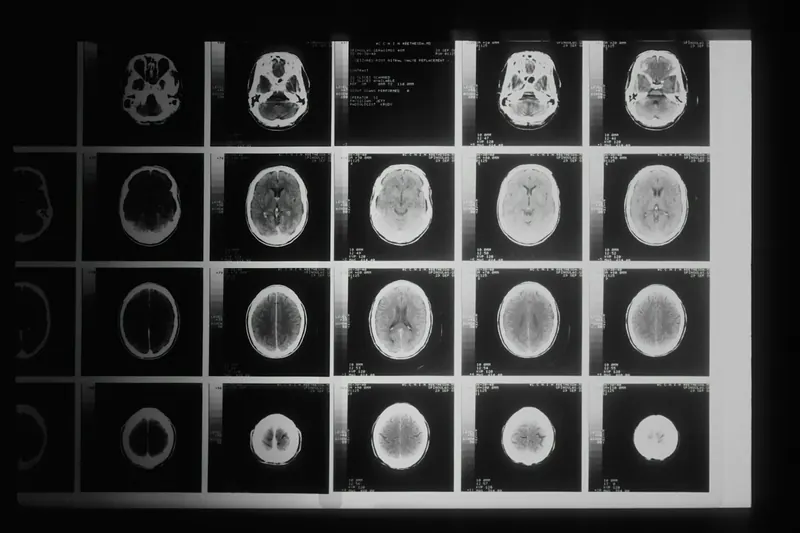
Before surgery, MRI and other scans are studied to plan the operation. You are given general anaesthesia and are fully asleep.
- 2
Your head is held still in a frame. A small area of hair may be shaved and the skin cut is planned to heal neatly.
- 3
The surgeon opens a small window in the skull (craniotomy) over or near the tumour. Guidance tools may help confirm the exact position.
- 4
Using the operating microscope, the surgeon removes as much of the tumour as is safe, while protecting important nerves and blood vessels. In some cases, part of the tumour is left behind on purpose to avoid harm to the brain.
- 5
The removed tissue is sent to the laboratory (histopathology) to find out exactly what type of tumour it is.
- 6
The bone is usually put back and fixed in place, and the skin is closed. You are then watched closely in the ICU (intensive care unit), often for a day or more.
Possible risks
Every operation carries some risk. Your surgeon will explain which of these apply to you.
Preparing for surgery
Recovery
After surgery, most patients spend some time in the ICU and then move to the ward. The hospital stay is often about 5 to 10 days, but it varies with the tumour, the operation and how you recover. Headache and tiredness are common for a few weeks.
Read moreShow less
The tissue report usually comes in about a week or two. Depending on the result, you may need no further treatment, regular scans, or radiotherapy and chemotherapy with a cancer specialist. Your surgeon will explain the report and the plan.
Some people need physiotherapy, speech therapy or help with daily activities for a while. Do not drive until your doctor says it is safe, especially if you had seizures. Keep all follow-up appointments and scans.
Questions patients ask
Is every brain tumour cancer?
No. Many brain tumours are benign (not cancer), such as most meningiomas. Even a benign tumour can cause problems if it presses on the brain. The exact type is known only after the tissue is tested.
Does every brain tumour need surgery?
No. Some small tumours that cause no symptoms can be watched with regular MRI scans. Some are treated with radiation or medicines. The decision depends on the tumour type, size, position and your general health.
Will my head be fully shaved?
Usually not. Often only a small strip of hair along the planned cut is shaved, and the hair grows back over the scar.
How long will I stay in hospital?
Often about 5 to 10 days, including a short stay in the ICU. Some patients go home sooner and some need longer, depending on how they recover.
Will the tumour come back?
It depends on the type of tumour and how much could be safely removed. Some benign tumours rarely come back once fully removed, while others need further treatment and regular scans. Your surgeon will explain this once the tissue report is ready.
Who can I talk to about my MRI report?
A neurosurgeon can explain what the scan shows and the choices available, after going through your reports.
Disclaimer: This information is for general awareness and is not a substitute for a medical consultation. Read moreSource: Content on this website was prepared with the help of AI tools (AI-assisted) and is medically reviewed by Dr. Kshitiz Nigam, MBBS, MS, MCh (Neurosurgery), Reg. No. MP-33282.